
A very personal reflection on motherhood as a podcast – my project, my own experience of being a mother, and a big thank you to my own mum!

A very personal reflection on motherhood as a podcast – my project, my own experience of being a mother, and a big thank you to my own mum!
In the course of our new project ‘Unlocking the secrets of cremated human remains’, we had the opportunity to CT scan recently excavated urns from St. Pölten. Now, why would one want to do this? There surely is not much to do for the patients in the urns!

In archaeological practice, Bronze Age urn burials are rarely found intact when they are excavated. Ploughing or other, later disturbance of the burial grounds has often destroyed the upper part of the burial. During rescue excavations, there is rarely the time to document all pieces of cremated remains, and urns are frequently emptied to get the remains to the osteologist for analysis. However, this case was different. St. Pölten’s city archaeologist Ronald Risy offered us two recently excavated urns excavated en bloc. This means that the entire urn, including the surrounding soil, had been wrapped in cling film and plaster for stabilization and recovered as a whole. This is the only way to preserve the whole context – the exact location of all cremated bone fragments, their position relative to each other, to grave goods within the urn, and to the filling material.

A detailed analysis of this contextual information can answer a number of questions about the burial ritual. How was the urn filled? Did Bronze Age people recognize the individual bones and place them in a specific anatomical order? Did they select specific parts of the body? Did they include charcoal and ash from the funerary pyre? Which dress elements and tools were cremated with the body and placed alongside the cremated remains? Did Bronze Age people fill the remaining space of the urn with pebbles or soil? Which taphonomic processes acted on the urns after burial?
It is a well-known truth that all excavations are destructive – they destroy the original layers, contexts and sequence of deposition despite archaeologists’ best efforts to record all possible information. It is therefore a good idea to document the burial context of the urn in as much detail as possible, and CT scans are an amazing tool to achieve the best possible visual documentation. Thanks to Fabian Kanz and his colleagues, we got an out-of-hours appointment at the Dental Clinic of the Medical University of Vienna.

The images and 3D models from CT scans cannot only guide the micro-excavation (it is always good to know what to expect!), but also help with a typical problem of analyzing cremated remains: the bones fragment further when they are excavated and handled, to the point where measurements are no longer possible. Measurements, however, are particularly important for estimating biological sex and body height. The CT scans enable us to measure the bones in situ from the images. In addition, trabecular bone is often stabilized within the urn, but falls apart when taken out. Pelvic bones, femoral heads, and other characteristic bones can be identified and in part already analyzed through the images.
The CT scans help us select the best possible strategy to disentangle soil, cremated remains and pottery. We can clearly see the location of each pottery fragment, the extent to which the urns are fragmented and how they collapsed from soil pressure. Since our focus is on getting the optimal information from cremated remains, we will compromise on the preservation of pottery if necessary. One of the urns is almost intact, the other quite fragmented. It will be exciting to take them out of their cast!
Check out a video of one of the CT scans here:
Acute or chronic diseases of the upper and lower respiratory tracts have always affected people’s everyday lives – in the past and today. They include colds, influenza, tonsillitis, sinusitis, bronchitis and chronic bronchitis, pleurisy, pneumonia, asthma and lung cancer. Virus-related diseases such as Covid-19 have been emerging sporadically for millennia. Environmental factors, as well as working and living conditions in settlements play a role in their development and spread. Air pollution and poor air quality support the emergence of respiratory diseases.
Infectious diseases spread by breathing in particles that an infected person coughs or sneezes, by touching body fluids directly or surfaces contaminated with saliva, blood, sweat or secretions. Fever, cough, breathlessness, sore throat, fatigue, chest pain while breathing or coughing, as well as muscular pain and headache may accompany respiratory diseases.
Most of these symptoms primarily affect soft tissues, but after persisting for some months, they manifest as discrete changes on the bone surface of skeletal remains. These traces can be investigated in the skeletal remains from archaeological contexts. Most common signs of respiratory diseases are found at the nasal floor and cavity (rhinitis), the paranasal sinuses (sinusitis) and the ribs (pleurisy). Especially pleurisy, the inflammation of the pleura that separate the lungs from the chest wall, leaves traces on bones due to the direct contact of the tissue with the ribs. Normal bone surfaces appear smooth, whereas porosities, vessel imprints, or plate-like new bone formations indicate pathological changes. We can differentiate active and healed lesions, but there are also mixed examples (see figure).
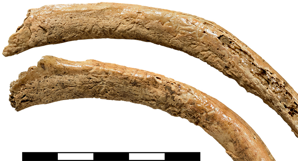
Partially healed pleurisy lesions at the internal side of the ribs of a 16-20-year-old female individual from Early Bronze Age Schleinbach, Lower Austria. © NHM Wien, W. Reichmann
Medical treatment is available for many respiratory diseases today. However, not all people have access to necessary medicines. First historical reports of flu epidemics date back to Ancient Greece, but outbreaks of flu and other viral diseases can probably be assumed for much earlier times. Without understanding the underlying causes, the diseases were difficult to treat. Left untreated, some were life-threatening or deadly, especially for undernourished people or individuals with a weakened immune system.
References
Gresky, J., and Schultz, M. 2011. “Einflüsse von Klima- und Wohnbedingungen auf Erkrankungen der Nasennebenhöhlen am Beispiel der population des bajuwarischen Gräberfeldes von Hartig (Oberpfalz),” in N. Benecke and S. Flohr (eds) Beiträge zur Archäozoologie und Prähistorischen Anthropologie 8. 83–94. Langenweißbach: Beier & Beran.
Klingner, S. 2016. Ätiologie und Epidemiologie der Erkrankungen des Respirationstraktes im Frühneolithikum Mitteleuropas am Beispiel der linearbandkeramischen Population von Wandersleben. PhD Thesis, Universität Leipzig.
Pany-Kucera, D., Berner, M., Reschreiter, H., Kern, A., and Kowarik, K. 2018. “Chronische Entzündungen der Nasennebenhöhlen als Hinweis auf die Umweltbedingungen im eisenzeitlichen Hallstatt,” in J. Drauschke et al. (eds) Lebenswelten zwischen Archäologie und Geschichte. Festschrift für Falko Daim zu seinem 65. Geburtstag, Monographien des RGZM 150. 985–995. Mainz: Verlag des Römisch-Germanischen Zentralmuseums.
Roberts, C. A. 2007. A Bioarchaeological Study of Maxillary Sinusitis. American Journal of Physical Anthropology 133: 792-80.
In the middle of a pandemic, we are constantly reminded to wash our hands. That handwashing is effective to stop the spread of disease, however, has not been common knowledge for more than a few centuries. This is a tribute to an early hero of handwashing and hospital hygiene: Ignác Semmelweis. Despite the fact that bacterial causes of infection were unknown at his time, he saved the lives of numerous mothers by advocating handwashing on the maternity ward.

Ignaz Semmelweis, aged 42 in 1860,
copperplate engraving by Jenő Doby
Ignác Semmelweis was born in Budapest, Hungary, in 1818. His birthplace, the Semmelweis Museum of Medical History, is a place well-worth visiting if you like quirky, small museums with astonishing collections.
Semmelweis moved to Vienna in 1837 to study law, but soon switched to medicine and was awarded his doctorate in 1844. He took up a position in the obstetric department of the General Hospital in Vienna, which was divided in two clinics: doctors and medical students worked in the first clinic, whereas midwifes were trained in the second. The mortality rate of women giving birth in the first clinic was between 5 and 15%; far fewer women died of puerperal fever in the second.
The reasons for this were unknown – germ theory had not yet been formulated. Doctors and students routinely performed clinical sections on patients that had died of child bed fever and then went on to examine women in labour. It was only after one of Semmelweis friends died of sepsis after accidentally being cut with a scalpel during a section that he made a crucial connection.
He discovered that the incidence of puerperal fever could be drastically reduced by disinfecting hands with chlorinated lime solution, not just between forensic investigations and examining patients, but also between each vaginal examination. Systematic hand hygiene lowered women’s mortality on the ward to 1.3 % in 1848.
His landmark study „Die Ätiologie, der Begriff und die Prophylaxe des Kindbettfiebers“ (Etiology, concept and prophylaxis of child bed fever) and his tireless avocation of hand washing was not met with enthusiasm by his contemporaries. Evidence-based medicine was yet to become established, hygiene was considered a waste of time and doctors believed that gentlemen’s hands could not transmit disease.
The tendency to reject new evidence or new knowledge because it contradicts established norms, beliefs, or paradigms has been named ‘Semmelweis reflex’ after the reaction of some of his colleagues. Semmelweis must have been a controversial, quarrelsome character, who attacked his critics in a series of open letters, in which he went as far as to accuse them of murder. Perhaps this serves as a reminder that the right way of dissemination is almost as important as the research results themselves!
Only after his early and obscure death in an insane asylum and the discovery of bacteria as the cause of infections of the female reproductive tract, Semmelweis became famous as the ‘Saviour of Mothers’. The unveiling of a new statue by the Hungarian artist Péter Párkányi Raab at the campus of the Medical University of Vienna marked his recent 200th birthday a gift from the Semmelweis University Budapest.

Links
https://en.wikipedia.org/wiki/Ignaz_Semmelweis
https://www.pastmedicalhistory.co.uk/ignaz-semmelweis-the-saviour-of-mothers/
Last year, my team and I have participated in filming a documentary on gender (pre-)history in the Natural History Museum in Vienna. It was an exciting, interesting day. We answered a number of interview questions on motherhood in prehistory and the role of women in society. We were filmed walking around the museum and handling artefacts. I have not seen the end product yet, but here are the broadcasting dates (in German):
Geschlechterkonflikt – Frauenbilder der Geschichte
52 min. TV Dokumentation auf ARTE
07. März 2020 um 21:05 Uhr
TERRA X: Mächtige Männer – Ohnmächtige Frauen? Neue Fakten aus der Vergangenheit
43 min. TV Dokumentation auf ZDF
21. Juni 2020 um 19:30 Uhr

This gallery contains 10 photos.
Originally posted on Archaeo𝔡𝔢𝔞𝔱𝔥:
Having reviewed my keynote lecture at the 1st CRUMBEL workshop in Brussels on Wednesday 16th October (and check out my Twitter Moment here), I want to briefly review the subsequent 2 days of presentations and posters on…
via Early Europeans bottle-fed babies with animal milk — The Bioarchaeology of Childhood | Sian Halcrow
written by Doris Pany-Kucera, 12.9.2019 in Vienna
Physical anthropologists have long desired to assess past pregnancies and births on human skeletal remains. They developed different methods, building on features of the pelvis that differ between male and female skeletons. However, none of these features, e.g. the preauricular sulcus or dorsal pubic pitting, are always present on the pelvic bones after partuition. To this day, the topic remains hotly debated.
In the scope of our project, which aims to link women’s reproductive and social status in prehistoric societies, we combine context information from graves such as grave goods or grave depth with skeletal data. We therefore record a set of pelvic features on the skeleton. Some of the observed features like the preauricular sulcus seem to relate to the female sex. The so-called ‘true’ preauricular sulcus, a very deep groove at the ilium bone next to the sacroiliac joint, for instance, only occurs in females in this shape. The question that remains to be answered is why this is the case.
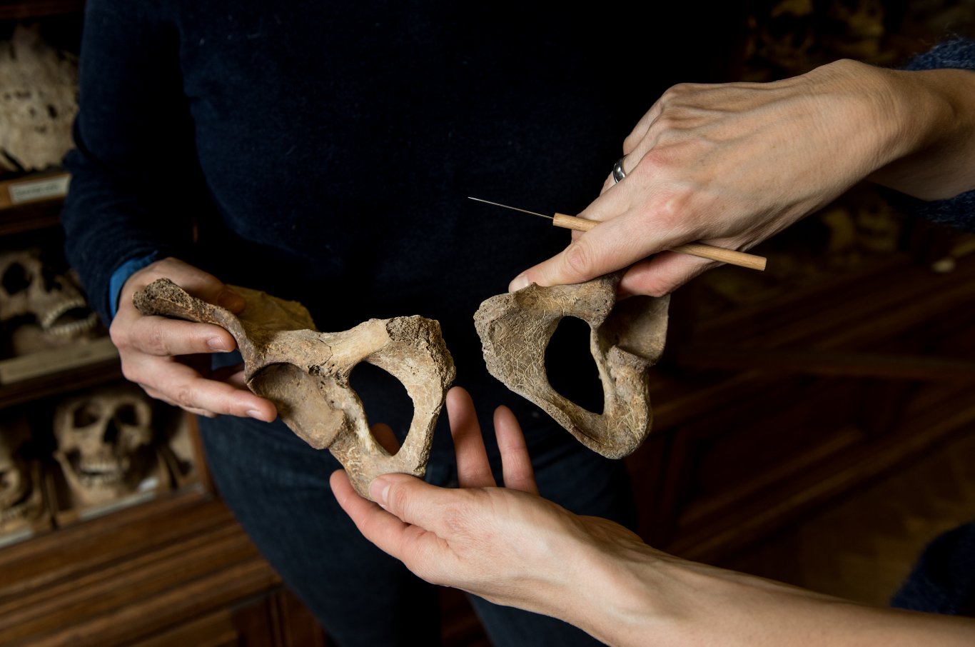
A Bronze Age female pelvis under scrutiny. Photo: Luiza Puiu
The adaptation of the female pelvis to cope with childbearing begins in adolescence. In the Austrian Bronze Age, archaeological evidence indicates first births at an early age. We therefore also included adolescent females in our analysis. During the data collection, we noticed a large, bilateral bony extension in the front section of a sacrum, next to the auricular facet of the sacroiliac joint, in a female skeleton for the first time. The extension was hard to interpret, as it was not a simple arthrosis. It appeared to be something special, and we began to pay special attention on local modifications in this area. In the course of gathering further data, we found another change at this location in the shape of a notch; interestingly, it was in a juvenile female individual. From then onwards, we found both changes more frequently and recorded them systematically in our skeletal series. We decided to term them sacral preauricular extension and sacral preauricular notch and recently published our findings.
The scientific hypothesis to explain the described changes is that they stand in a causal relationship with pregnancy and birth events. We came to this interpretation on the basis of our results. First, we so far found the changes only in female skeletons aged 16 or over, and we have meanwhile analyzed more than 400 male and female individuals. Second, we also found the same changes in a so-called identified collection, where biological and socioeconomic data are available: they only occur in females with two or more children.
We believe that the formation of the sacral preauricular extension forms through increased compressional forces at this front section of the sacrum, most likely in the course of traumatic birth events. This includes deliveries with complications, such as prolonged labour – when the child’s head only barely fits through the mother’s pelvis. Additional mechanisms include that the hormonally induced increased pelvic joint mobility often leads to a postural change during pregnancy, and together with the weight gain contributes to the formation of the feature.
Furthermore, we suggest that the sacral preauricular notch arises from pregnancies and births at a young age, as we found the ossifying epiphysis at the sacrum to be affected in adolescents. Perhaps early physical work and early pregnancy may both explain at least the sacral preauricular notch, but we did not find distinct activity-related changes in the analyzed skeletons. We are therefore confident that the changes relate to pregnancies and births. The frequency of the bony modifications varied between the analyzed sites, which may partly be a result of preservation issues, but may point to different birthing practices or birth spacing.
In general, the interpretation of pelvic features in relation to pregnancy and parturition is still difficult, because we know little on the process of their formation. Many factors such as age, weight, stature, biomechanical and musculoskeletal conditions, pelvic dimensions, genetic disposition, hormonal influences, or workload may influence if they occur and what exactly they look like. To move the discussion forward, we are working closely together with anatomists, radiologists and pelvic floor experts of the Medical University and General Hospital of Vienna.
On 28 November 2019, the project team organizes a workshop on female pelvic features with national and international experts from different professional backgrounds in the Natural History Museum in Vienna. Book your space if you are interested!
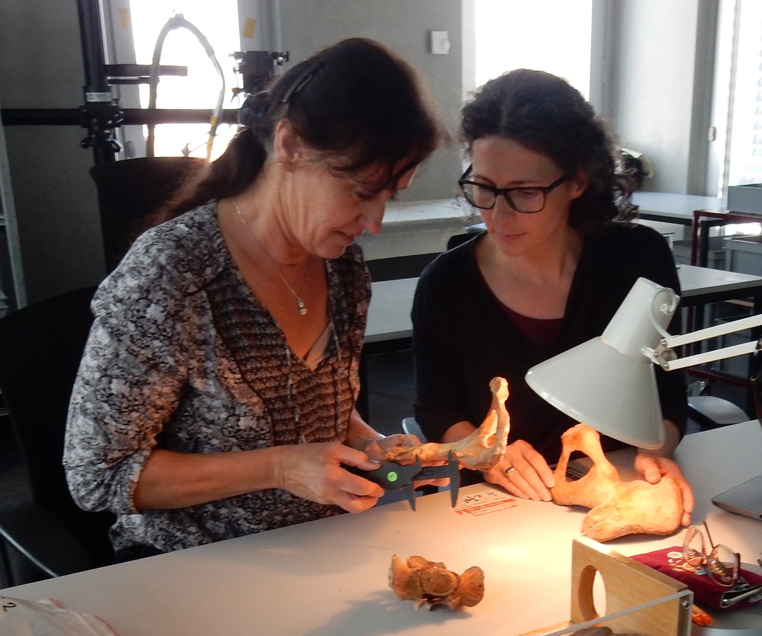
Michaela Spannagl-Steiner and Doris Pany-Kucera working on pelves
References
Pany-Kucera, D., M. Spannagl-Steiner, S. Argeny, B. Maurer-Gesek, W. J. Weninger, and K. Rebay-Salisbury. 2019. Sacral preauricular extensions, notches and corresponding iliac changes: new terms and the proposal of a recording system. International Journal of Osteoarchaeology: https://onlinelibrary.wiley.com/doi/epdf/10.1002/oa.2814.
Pany-Kucera, D., M. Spannagl-Steiner, W. Parson, B. Rendl, C. Strobl, L. Waltenberger, and K. Rebay-Salisbury. accepted. Motherhood, Social Relations and Violence at Schleinbach, Lower Austria: re-examining and assessing the Early Bronze Age human remains Archaeologia Austriaca.
Rebay-Salisbury, K., D. Pany-Kucera, M. Spannagl-Steiner, F. Kanz, P. Galeta, M. Teschler-Nicola, and R. B. Salisbury. 2018. Motherhood at early Bronze Age Unterhautzenthal, Lower Austria. Archaeologia Austriaca 102: 71-134.
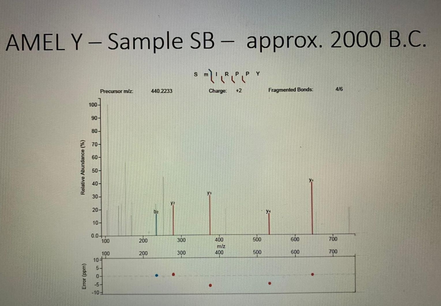
A new method of sexing juvenile human remains has recently been described in the literature (Parker et al. 2019, Stewart et al. 2017, Stewart et al. 2016), and it sounds almost too good to be true: sexually dimorphic amelogenin protein fragments can be identified in human enamel using high performance liquid chromatography.
Based on morphology alone, it is notoriously difficult to assign sex to children, as sexual dimorphism develops primarily after puberty. Osteologists therefore do not normally assign sex to children and adolescents under the age of about 16 to 18 (Cunningham, Scheuer, and Black 2016). In recent years, DNA analysis has been employed to determine the genetic sex of skeletons, but DNA analysis is destructive, its success depends on the preservation of the nuclear DNA and the costs are still prohibitive. The identification of peptides in tooth enamel, in contrast, is almost non-destructive – it only needs an incredibly small amount of tooth enamel gained by acid etching of a small area of the surface of the tested tooth. The tested area is hardy visible to the naked eye (wearing contact lenses, I could not see it).
As an archaeologists interested in age and gender, this is like a (slightly macabre) dream come true. We can finally answer a number of questions on sex-specific mortality patterns, on sex-preferences, and demography. Why is knowing the sex of buried children so important? We will be able to know if sex selection took place after birth and whether infanticide affected more girls or more boys. We can ask whether girls and boys were treated equally as babies and small children, for example in terms of access to food. We can investigate if children of both sexes were afforded the same burial rites. We will be able to tell if the sex of babies and infants was important, or if societies only responded to the differences between girls and boys later, as children matured. We will be much better able to understand how children ‘learn gender’, at what age girls and boys were socially recognized and treated as adolescents and adults. In summary, we can learn a lot about value systems linked to gender, about power relationships between the sexes, and about how they developed in past societies.
Department of Analytical Chemistry, University in Vienna
It is a lucky coincidence that Fabian Kanz, my collaboration partner in Forensic Medicine, has good contacts to the Department of Analytical Chemistry of the University in Vienna, where nanoLCMS/MS machines are part of the routine analytical equipment. Thanks to the efforts of Lukas Janker and Dina Schuster, a test on a series of modern deciduous and permanent teeth from individuals with known sex was successful, a laboratory protocol was established, and work on prehistoric teeth could begin. The first sex identification of a 5-6-year-old Bronze Age child via peptides in tooth enamel produced unambiguous results, as AMELY was clearly present. It’s a boy!
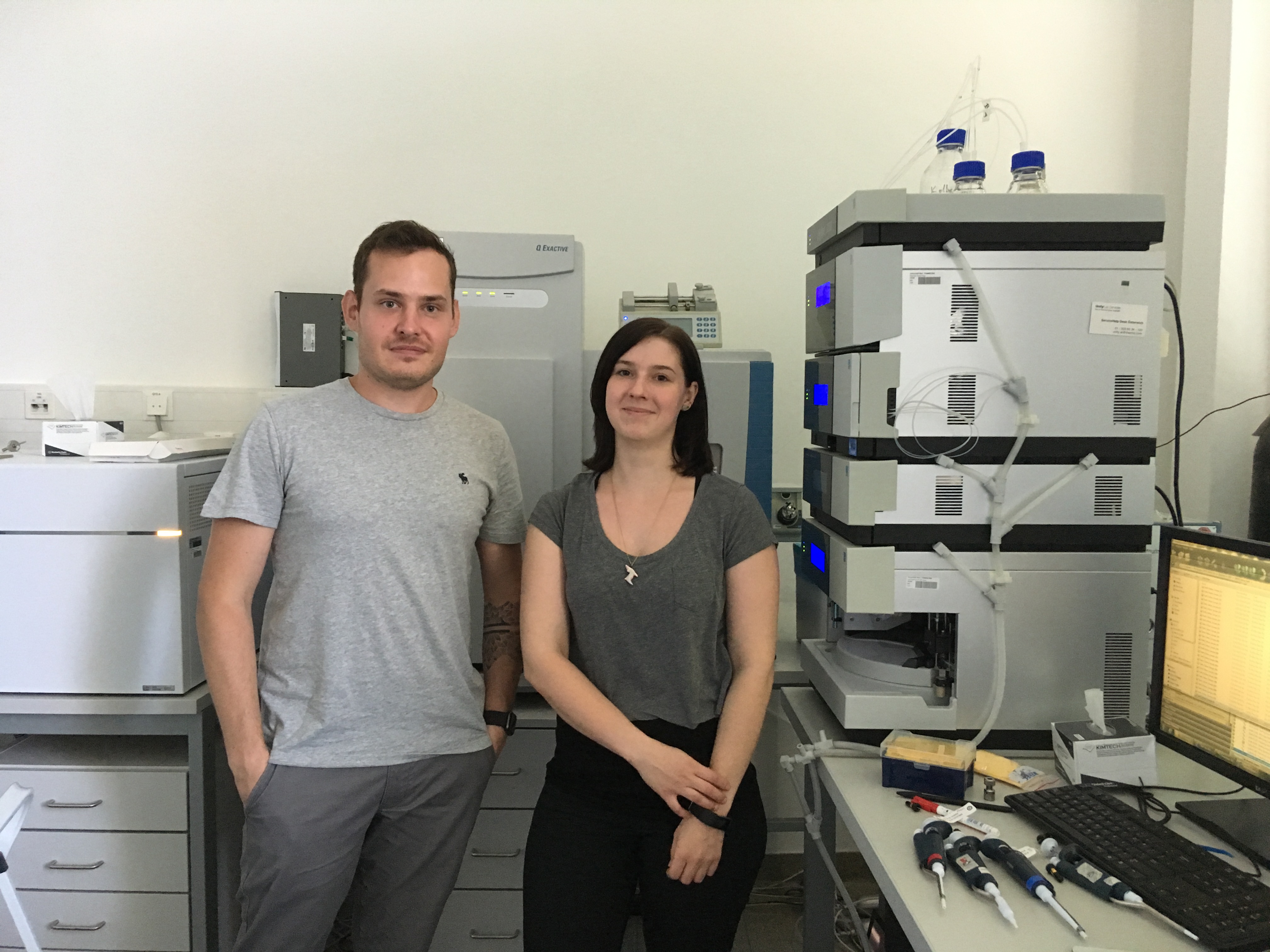
Lukas Janker and Dina Schuster in front of the QExactive orbitrap mass spectrometer
References
Cunningham, C., L. Scheuer, and S. Black. 2016. Developmental Juvenile Osteology, 2nd edition. London: Elsevier Academic.
Parker, G. J., J. M. Yip, J. W. Eerkens, M. Salemi, B. Durbin-Johnson, C. Kiesow, R. Haas, J. E. Buikstra, H. Klaus, L. A. Regan, D. M. Rocke, and B. S. Phinney. 2019. Sex estimation using sexually dimorphic amelogenin protein fragments in human enamel. Journal of Archaeological Science 101: 169-180.
Stewart, N. A., R. F. Gerlach, R. L. Gowland, K. J. Gron, and J. Montgomery. 2017. Sex determination of human remains from peptides in tooth enamel. Proceedings of the National Academy of Sciences.
Stewart, N. A., G. F. Molina, J. P. Mardegan Issa, N. A. Yates, M. Sosovicka, A. R. Vieira, S. R. P. Line, J. Montgomery, and R. F. Gerlach. 2016. The identification of peptides by nanoLC-MS/MS from human surface tooth enamel following a simple acid etch extraction. RSC Advances 6, 66: 61673-61679.
Recently, the results of our DNA analysis from the early Bronze Age site Schleinbach came in. We have been working on Schleinbach a bit longer than intended. It is an exciting site, as it includes single graves, a double and multiple burial, several individuals buried or deposited in former storage pits, and can tell us a lot about social relations and social stratification the Bronze Age. Many individuals have healed or perimortal fractures that suggest interpersonal violence.
We sent samples from two of the most interesting contexts to the Legal Medicine Department in Innsbruck, to test for maternal relationships between the buried individuals. Mitochondrial DNA is inherited only from the mother. Because it is present in many more copies per cell than nuclear DNA, mtDNA is more likely to be preserved in archaeological samples. Normally, offspring inherit an identical copy of the mother’s mtDNA, but random changes sometimes occur, which are then passed down the generations. Specific mutations characterize a haplogroup, a genetic group of people who share a common ancestor. MtDNA only shows a part of a person’s genetic history – the maternal linage.
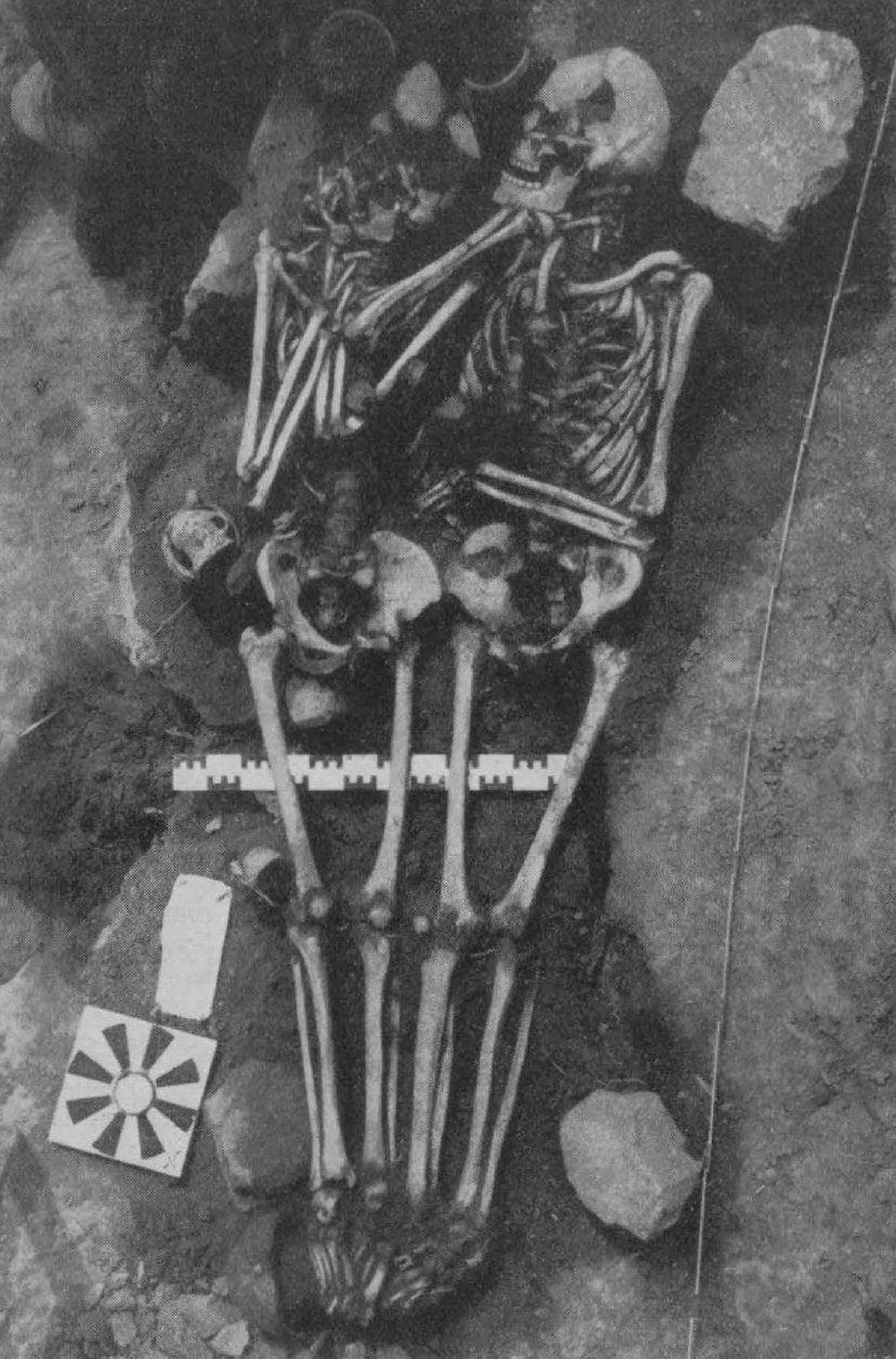
Double burial 30/31 from Schleinbach (Foto: Karl Kriegler, 1927)
The double burial 30/31 includes two male individuals who died at the ages of 27-30 and 30-35 years. Both had very similar perimortem fractures of the skull, which likely led to their death, and they were placed very closely together in a single grave. The bones of the feet overlap and the pelvic bones almost touch, giving rise to the suspicion that they were bound or wrapped together after death. It is unclear what happened to the men – perhaps they were executed by their own community, perhaps they died raiding a neighbouring village and were sent home dead and tied together, perhaps they died defending their own home during a surprise attack – we do not know. We only know they met a similar violent fate.
MtDNA analysis showed that the individuals did not only share the same haplogroup, the mitrochondrial haplotype was also identical, i.e. they share the same specific DNA sequences inherited together. The ‘brothers in arms’ were closely maternally related; perhaps they were indeed brothers, although we cannot exclude they were cousins or otherwise related.
Four individuals – an adult man and three children aged 3-4, 8-9 and 12 – were found deposited in a former storage pit at Schleinbach. Again, perimortal traumata suggest that at least one of the individuals met a gruesome fate, but it looks like all four died in quick succession. Only the two younger children, however, share the same mitotype and perhaps had the same mother. The 12-year-old was not their maternal sibling, and the adult man had his own haplogroup. It is thinkable that he was the father of the children.
In total, we had four different mitochondrial haplogroups in this sample – and of course the first thing to do is google them! The 12-year-old from the pit turns out to have J1c2 – a haplogroup shared with the famous King Richard III. He was king of England from 1483 to 1485, when he died in the Battle of Bosworth. Immortalized by William Shakespeare as a villain, his skeletal remains were rediscovered under a concrete car park in Leicester in 2012. Having lived in Leicester for five years, I got overly excited by the match.
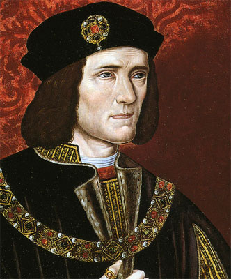
Richard III (Wikimedia Commons)
Is it possible that we found Richard III’s maternal ancestor in a Bronze Age grave in Austria? There are many generations between them. Our 12-year-old lived between 1906 and 1743 cal BC, as radiocarbon dating has revealed, and assuming 30 years for a generation, this would be about 110 generations between them. Many grand-grand-grand-grand-grands to write.
Turi King from the University of Leicester, who led the DNA analysis of Richard III, and Walther Parson found out Richard III belongs to a relatively uncommon subclade J1c2c3, and there isn’t a perfect match between the individuals. The two were definitely related, but perhaps not as closely as to be of any significance. The most recent common ancestor for our Bronze Age individual and Richard III might have lived 7800 to 11800 years ago – this is the assumed age of the branch J1c2. It is estimated that today, about 17 million people worldwide share J1c2 (Behar et al 2012, Logan and Brinkman 2017).
Literature
Behar, Doron M., M. van Oven, S. Rosset, M. Metspalu, E.-L. Loogväli, Nuno M. Silva, T. Kivisild, A. Torroni, and R. Villems. 2012. Reassessment of the Human Mitochondrial DNA Tree from its Root. The American Journal of Human Genetics 90, 4: 675-684.
Buckley, R., M. Morris, J. Appleby, T. King, D. O’Sullivan, and L. Foxhall. 2013. ‘The king in the car park’: new light on the death and burial of Richard III in the Grey Friars church, Leicester, in 1485. Antiquity 87: 519–538.
King, T. E., G. G. Fortes, P. Balaresque, M. G. Thomas, D. Balding, P. M. Delser, R. Neumann, W. Parson, M. Knapp, S. Walsh, L. Tonasso, J. Holt, M. Kayser, J. Appleby, P. Forster, D. Ekserdjian, M. Hofreiter, and K. Schürer. 2014. Identification of the remains of King Richard III. Nature Communications 5: 5631.
Logan, I. S., and D. N. Brinkman. 2017. King Richard III and his mitochondrial DNA haplogroup J1c2c3. The Journal of Genealogy and Family History 1, 1: 1-14.
Rebay-Salisbury, K. 2018. “Vielversprechende Ansätze und kleine Irrwege: die Interpretationsgeschichte frühbronzezeitlicher Bestattungen am Beispiel Schleinbach,” in F. Pieler and P. Trebsche (eds) Beiträge zum Tag der Niederösterreichischen Landesarchäologie 2018. 45-56. Asparn: Niederösterreichisches Landesmuseum.